SONICC imaging, which includes both SHG and UV-TPEF imaging methods, produces black and white images that makes protein crystal detection easy. (For more information about how SONICC imaging works, see SONICC Imaging.)
While this topic goes over all of the main components, the SONICC Benchtop ROCK IMAGER hardware component guide has also been made into an interactive video. See our online SONICC demo to virtually open and explore the insides of SONICC Benchtop.

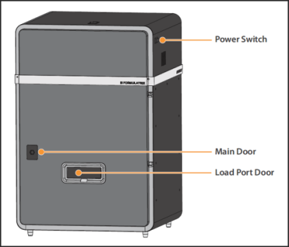
SONICC External Components
The power switch is located on the right-hand side of the SONICC benchtop imager.
The main door is latched securely with a hex key.
Plates can be safely added to SONICC while the door is closed and latched by placing a plate in the load port door.

SONICC Internal Components
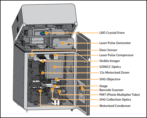
In UV-TPEF mode, the crystal oven controls the LBO crystal at a precise temperature to maximize a green femtosecond pulse laser output. Green ultra-short laser pulses produce an ultraviolet two photon excited florescent signal on the amino acid tryptophan.
A fiber-based femtosecond laser pulse generator provides a robust laser power source. Laser controls are fully integrated with the ROCK IMAGER software, removing the need for manual adjustments.
The laser will shut off if the door sensor detects that the door is open.
The laser pulse compressor produces a laser pulse length for less than 200 femtoseconds. The ultra-short laser pulse is necessary for second harmonic generation.
The 5 megapixel, 2/3" color CCD camera captures high-quality images at up to 25 frames per second.
The novel design integrates SHG, UV-TPEF, and laser scanning in an ultra-compact footprint.
The 12x continuous zoom delivers a maximum field of view of 9.49 mm for resolvable features as small as 0.83 microns.
The SHG objective provides a 2.3 mm field of view, which is enough to cover most SBS standard wells without the need for image stitching.
The SONICC stage provides precise plate manipulation to within 5 microns.
The on-the-fly barcode scanner identifies plates loaded into SONICC by reading the barcode label and interpreting the label's information.
The PMT collects the SHG and UV-TPEF signals and converts them to electronic signals.
The transmission collection optics collect SHG signal while the epi collection optics collect the UV-TPEF signal.
The motorized condenser provides high-contrast images with no sample heating to assist with drop location.

|
|
| RIC-V38R119 |